What Are the Top Types of Specimen Tubes?
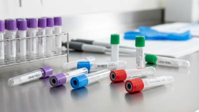
Choosing the right specimen tube is a small decision with visible consequences: it can affect sample handling, test compatibility, and the reliability of laboratory results. A lavender-top tube sits beside a gold-top tube on the collection tray, but their additives and intended uses are not interchangeable. Clinical chemist Dr. Guido F. Lippi’s work on preanalytical quality highlights a central principle: specimen quality begins before analysis. That is a paraphrase of his research, not a verbatim quotation.
This guide explores common tube types, including serum, plasma, anticoagulant, and specialty tubes. It explains how cap color, additive, fill volume, and mixing instructions help distinguish them. Color is a useful cue, not a universal guarantee; conventions can vary by manufacturer and region. Always check the product labeling and the laboratory’s collection instructions.
Details matter. An underfilled tube, a missed inversion, or a mismatched additive can complicate a sample’s journey. Not every error is obvious at the bedside. And no single tube fits every test. The sections ahead offer a practical overview for healthcare teams, laboratory learners, and anyone comparing specimen tube options. They do not replace local protocols or manufacturer guidance. Some distinctions may seem fussy. In specimen collection, they often are not.
What Specimen Tubes Are and How They Preserve Samples
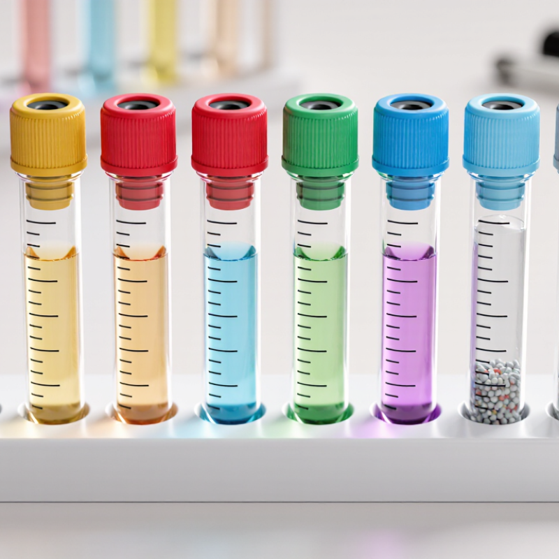
Specimen tubes are sterile, sealed containers designed to collect and transport samples while limiting contamination and changes before testing. For blood, an evacuated tube draws a measured volume through a needle. Its inner surface may contain an additive chosen for a specific test. Anticoagulants help keep blood from clotting; clot activators help serum form. Some tubes contain a gel that separates cells from liquid after centrifugation. Small details matter. A short fill, vigorous shaking, or delayed processing can affect results.
The tube preserves a sample by controlling contact, separation, and handling—not by stopping every change. Plebani’s review in Clinical Chemistry and Laboratory Medicine reported that 46–68.2% of laboratory errors occur in the preanalytical phase, which includes collection and transport. That range is a reminder: the tube is only one part of quality control. Follow the laboratory’s collection and storage instructions, since acceptable conditions vary by test. Keep the cap secure, mix additive tubes gently as directed, and note collection time when required. Tube-cap colors can help identify additives, but color conventions may vary. I would not rely on color alone.
How Tube Additives Determine Specimen Type
What Are the Top Types of Specimen Tubes?
How Tube Additives Determine Specimen Type
A tube’s additive helps determine whether a laboratory receives whole blood, plasma, or serum. Color can offer a quick clue, but cap colors are not universal. Check the label and local collection guide. Small details matter.
EDTA prevents clotting by binding calcium, so these tubes commonly provide whole blood for blood counts. Sodium citrate also binds calcium, but in a controlled ratio with blood. This makes it useful for many coagulation tests; the fill line matters because underfilling can alter that ratio. Heparin prevents clotting through a different mechanism and is often used when plasma is needed for chemistry testing. These are not interchangeable.
Serum tubes allow blood to clot before the liquid portion is separated. Some contain a clot activator, while others include a gel barrier to help separate serum from cells during processing. Fluoride-containing tubes can slow glycolysis, helping preserve glucose levels after collection. Timing and handling still matter. Additives guide the specimen type, but they do not guarantee a valid result on their own. I find this easy to overlook: the right tube, filled and handled incorrectly, may still produce an unsuitable specimen.
Common Serum and Clot-Activator Tubes
Serum tubes are used when testing requires serum, the liquid remaining after blood clots and cells are separated. Plain serum tubes contain no anticoagulant. Clot-activator tubes have a surface coating or additive that helps a clot form. Some also contain separator gel, which creates a barrier during centrifugation. Not every tube has gel. Check the tube’s instructions.
After collection, mix only as directed. Many clot-activator tubes require gentle inversions, but the exact handling can vary by design. Allow the specimen to clot fully before centrifugation; rushing may leave fibrin strands that complicate pipetting or interfere with analyzer probes. A clear serum layer is useful, but appearance alone cannot confirm suitability. Follow the laboratory’s validated timing and centrifuge settings. Small details matter.
Tips: Check the additive on the label, not cap color alone, since color conventions can vary. Keep tubes upright while clotting and note collection time when required. It is easy to assume an imperfect sample will still work. If a tube is underfilled, hemolyzed, or contains visible fibrin after separation, check local acceptance criteria rather than guessing.
What Are the Top Types of Specimen Tubes? – Common Serum and Clot-Activator Tubes
| Tube Type |
Typical Additive |
Specimen After Processing |
Common Uses |
Key Handling Notes |
| Plain red-top tube |
No additive in some glass tubes; plastic versions may contain a clot activator |
Serum |
Routine chemistry, serology, and other tests requiring serum, as specified by the laboratory |
Allow the blood to clot fully before centrifugation. Check the tube instructions because additives can vary. |
| Gold-top serum separator tube (SST) |
Clot activator and a polymer gel separator |
Serum separated from blood cells by a gel barrier after centrifugation |
Many routine chemistry and immunology tests |
Mix gently by inversion according to the tube instructions. Allow adequate clotting time before centrifugation. |
| Red-and-gray mottled-top serum separator tube |
Clot activator and a polymer gel separator |
Serum separated from blood cells by a gel barrier after centrifugation |
Routine serum testing, depending on laboratory requirements |
Often called a “tiger-top” tube. Confirm that the specific test accepts this tube type. |
| Orange-top rapid serum tube (RST) |
Thrombin-based clot activator and a gel separator |
Serum separated from blood cells by a gel barrier after centrifugation |
Selected tests requiring faster serum preparation |
Designed for rapid clot formation; follow the specified clotting and centrifugation instructions. |
Note: Tube colors and additives may vary by manufacturer and collection system. Follow the laboratory’s test requirements and the tube’s instructions for mixing, clotting, and centrifugation.
Common Plasma and Whole-Blood Collection Tubes
Plasma and whole-blood tubes are chosen according to the test, not just their cap color. Plasma comes from blood collected with an anticoagulant and then centrifuged. Whole blood stays unseparated, which helps preserve its cells for tests such as a complete blood count. Small details matter. The wrong additive can affect results.
EDTA tubes are commonly used for hematology because they help prevent clotting while preserving blood cells. Sodium citrate tubes are used for coagulation testing and require the correct fill volume; underfilling can alter the blood-to-additive ratio. Heparin tubes are used for certain chemistry tests. They produce plasma after centrifugation. Tube colors can vary, so check the local laboratory’s instructions rather than relying on color alone.
Tips: Confirm the test and tube before collection. Fill citrate tubes to the marked line, then gently invert tubes as directed; shaking can damage cells. Keep samples labeled and handle them promptly. One easy-to-miss point: “whole blood” describes the sample, not a single tube type. Follow the laboratory’s handling and transport requirements.
How to Match Specimen Tubes to Laboratory Tests
What Are the Top Types of Specimen Tubes?
How to Match Specimen Tubes to Laboratory Tests
Choosing a specimen tube starts with the test, not the color. A lavender-top tube usually contains EDTA and is commonly used for complete blood counts. Light blue tubes contain citrate and are used for many coagulation tests. They must be filled to the marked line, since the blood-to-additive ratio matters. Small details matter.
Serum tubes, often red or gold, are used for many chemistry and serology tests. Green-top tubes generally contain heparin and are common for plasma chemistry testing. Gray-top tubes are often selected for glucose testing because their additives help limit changes after collection. These are useful patterns, not universal rules. Tube colors and additives can vary by laboratory and region.
Check the laboratory’s test directory before collecting a sample. It identifies the required tube, specimen type, and any handling instructions, such as chilling or prompt separation. Follow the stated order of draw to reduce additive carryover, and gently invert tubes when instructed. Do not shake them. A tube may look correct yet contain the wrong additive or volume. When instructions seem unclear, pause and verify them; guessing can mean recollection and delayed results.

 +86 178 5514 5298
 CONTACT NUMBER
 CONTACT NUMBER